Research & Selected Publications
All publications on Google Scholar · * Equal contribution
2026 · IEEE Journal of Biomedical and Health Informatics
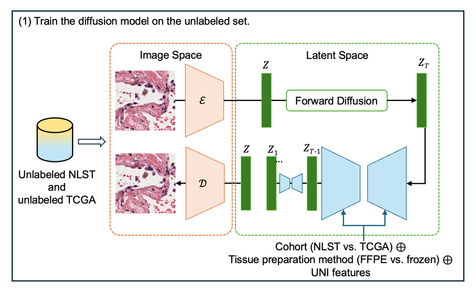
Semi-Supervised Domain Adaptation with Latent Diffusion for Pathology Image Classification
We use a latent diffusion model trained on unlabeled source and target images to generate pathology images that preserve tissue morphology while incorporating target-domain appearance. Adding these synthetic images to classifier training improved lung adenocarcinoma growth-pattern classification on a held-out target cohort, increasing weighted F1 from 0.611 to 0.706.
2025 · Journal of Biomedical Informatics, 167, 104833
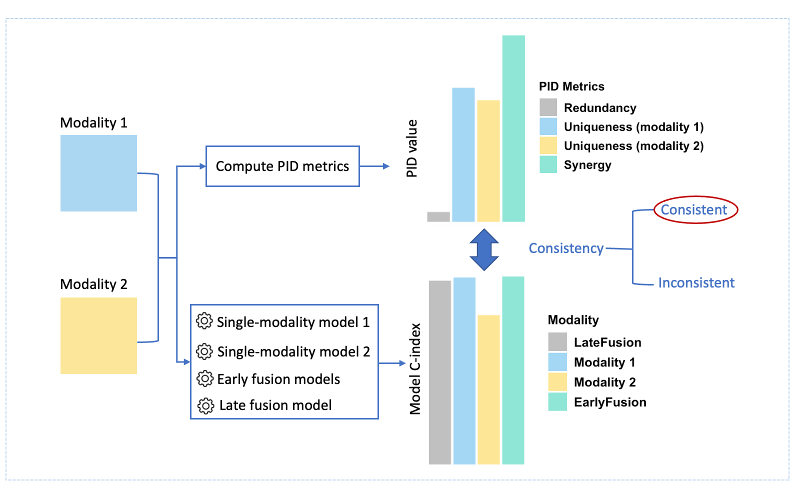
Evaluating an information theoretic approach for selecting multimodal data fusion methods
Can information theory tell us how to combine medical data? We evaluated partial information decomposition across seven modality pairs from four biomedical cohorts. The metrics did not consistently identify the best-performing fusion strategy; we investigated parameter selection and uncertainty estimation to improve their reliability.
2024 · SPIE Medical Imaging: Digital and Computational Pathology
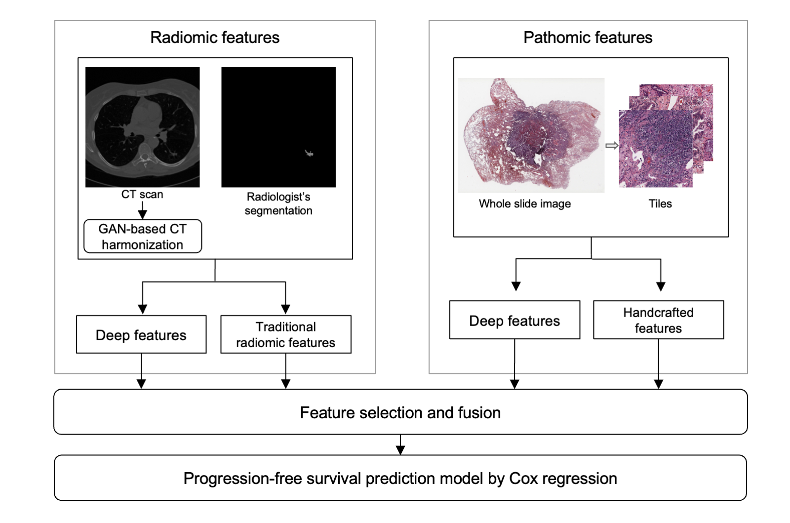
Using integrated radiomic and pathomic-based models to predict progression-free survival in early-stage lung adenocarcinoma
We combined features from CT scans and whole-slide pathology images to predict progression-free survival in 106 patients from the National Lung Screening Trial. With repeated five-fold cross-validation, the fused Cox model achieved a mean test C-index of 0.634, compared with 0.612 for radiomics alone and 0.584 for pathomics alone.
2024 · SPIE Medical Imaging: Computer-Aided Diagnosis
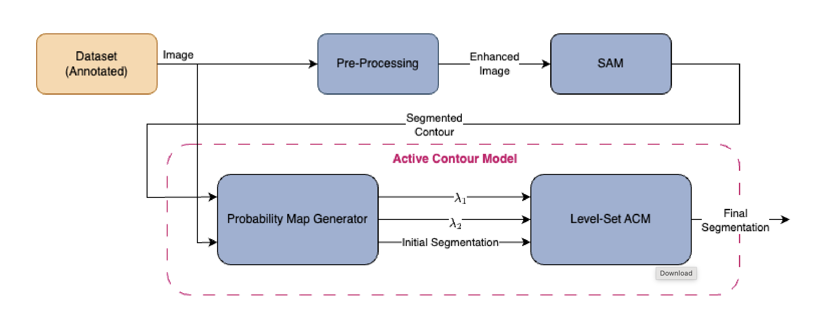
Refining Boundaries of the Segment Anything Model in Medical Images Using an Active Contour Model
We combine the Segment Anything Model with a level-set active contour model to refine medical image boundaries at test time. Evaluated on skin lesion and retinal vessel datasets, the approach addresses segmentation errors in images with low contrast or artifacts without additional supervised fine-tuning of SAM.